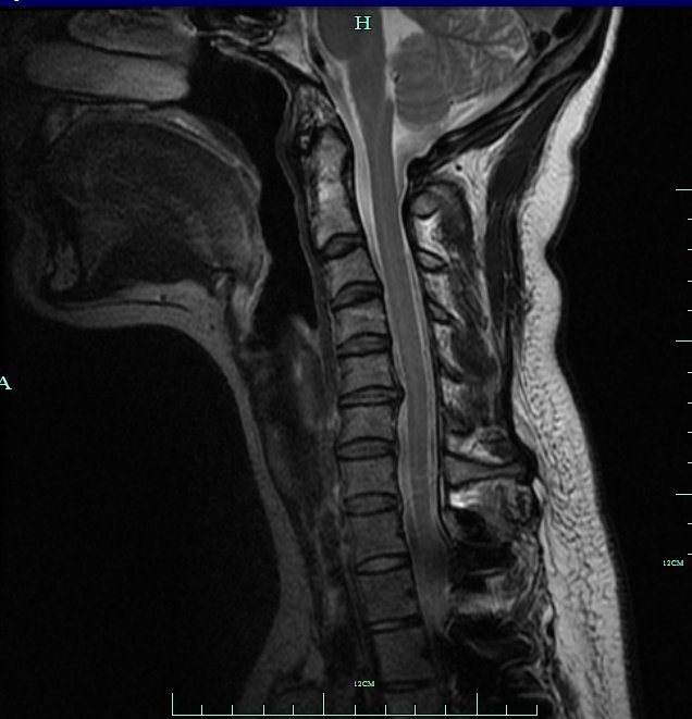
I have received the results from my MRI: There is NO CHANGE in size!!! The measurements for each part of my heart are literally exactly the same. This caused some confusion on my part, because the only reason I had the MRI in the first place was due to my echo showing aortic enlargement great enough to warrant an MRI. So why do the results contradict each other? Well, someone pointed out that echos have some flexibility in measurement and are not as accurate as MRIs are.
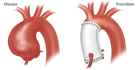
I cannot even begin to explain the amount of relief I feel. I know that I still have to have heart surgery down the road, but it is not yet the time to do so. This gives me time to try and manage my anxiety and become stronger and healthier (e.g. quit smoking) so that when I do have surgery, the recovery process will go much more smoothly.
There is still the issue of the pain in my left arm/chest. In addition, my constant brain fog/dizziness remains. For these issues, I have scheduled an appointment with my cardiologist, but the earliest they can see me is the end of September (a little over a month away). I take some comfort in thinking that if there was something significantly wrong, the MRI would have picked up on it. All I can do is wait for my appointment next month to see if other tests are needed, etc. Until then, I am going to try to relax a little bit; I have been so on edge since the news from my echo that I have been a complete wreck. I can also go ahead with next semesters classes (wasn't sure if I'd have to cancel them for surgery or not).
God is good. Life is beautiful. If all of my struggles have taught me anything, it is to appreciate what you have.
*RESULTS AS SEEN ON BRONSON'S WEBSITE*:
"HISTORY: thoracic aneurysm.
PROCEDURE: Magnetic resonance angiography, chest (excluding myocardium), with
or without contrast material(s)
TECHNIQUE: Without contrast 2-D time-of-flight with post-processing including
MIP and 2-D reconstructions was performed. Without contrast multiplanar
FIESTA sequences were performed. Without contrast cardiac gated double IR
sequences were performed.After injection of intravenous gadolinium contrast,
3-D MR angiography was performed, with post-processing to include MIP and 2-D
reconstructions.
Contrast: 20 mL Multihance
FINDINGS:
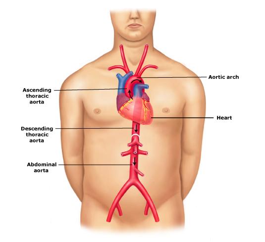
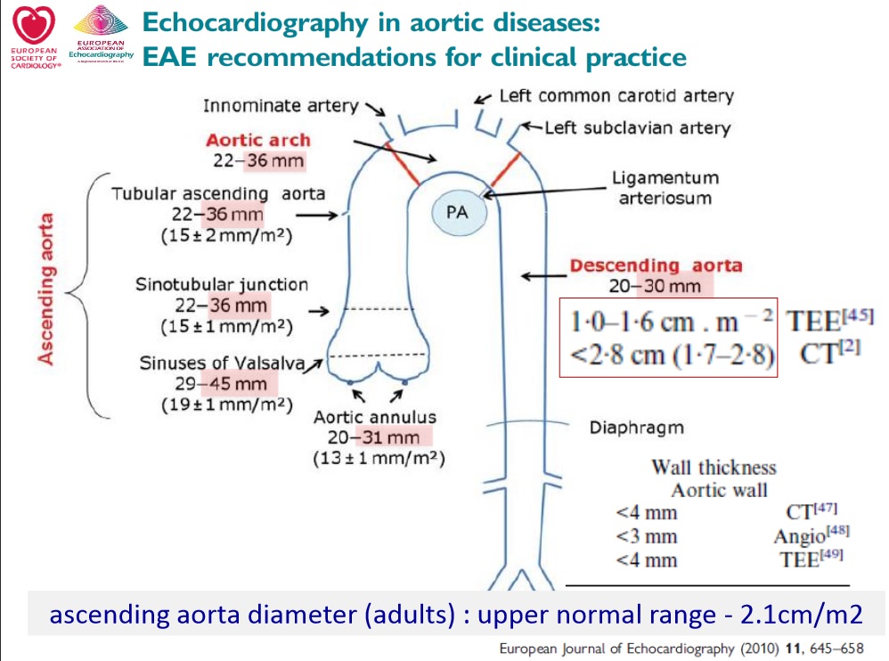
Comparison with 4/17/2013. Dilated ascending aorta again seen. There has been
no significant change. Measurements are identical to the previous exam.
Aortic annulus 2.2 x 2.8 cm
Sinuses of Valsalva 4.1-4.6 cm
Sinotubular junction 4.1 cm
Ascending aorta at RPA 3.1 cm
Ascending aorta at innominate 2.0 cm
Transverse distal arch 1.8 cm
Descending aorta at RPA 1.7 cm
Descending aorta at hiatus 1.7 cm
Aortic regurgitation again seen, not significantly changed from previous. No
other abnormality is seen. The visualized mediastinum and abdominal organs
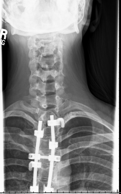
are unremarkable. Artifact from spine fixation hardware again noted."
I cannot even begin to explain the amount of relief I feel. I know that I still have to have heart surgery down the road, but it is not yet the time to do so. This gives me time to try and manage my anxiety and become stronger and healthier (e.g. quit smoking) so that when I do have surgery, the recovery process will go much more smoothly.
There is still the issue of the pain in my left arm/chest. In addition, my constant brain fog/dizziness remains. For these issues, I have scheduled an appointment with my cardiologist, but the earliest they can see me is the end of September (a little over a month away). I take some comfort in thinking that if there was something significantly wrong, the MRI would have picked up on it. All I can do is wait for my appointment next month to see if other tests are needed, etc. Until then, I am going to try to relax a little bit; I have been so on edge since the news from my echo that I have been a complete wreck. I can also go ahead with next semesters classes (wasn't sure if I'd have to cancel them for surgery or not).
God is good. Life is beautiful. If all of my struggles have taught me anything, it is to appreciate what you have.
*RESULTS AS SEEN ON BRONSON'S WEBSITE*:
"HISTORY: thoracic aneurysm.
PROCEDURE: Magnetic resonance angiography, chest (excluding myocardium), with
or without contrast material(s)
TECHNIQUE: Without contrast 2-D time-of-flight with post-processing including
MIP and 2-D reconstructions was performed. Without contrast multiplanar
FIESTA sequences were performed. Without contrast cardiac gated double IR
sequences were performed.After injection of intravenous gadolinium contrast,
3-D MR angiography was performed, with post-processing to include MIP and 2-D
reconstructions.
Contrast: 20 mL Multihance
FINDINGS:
Comparison with 4/17/2013. Dilated ascending aorta again seen. There has been
no significant change. Measurements are identical to the previous exam.
Aortic annulus 2.2 x 2.8 cm
Sinuses of Valsalva 4.1-4.6 cm
Sinotubular junction 4.1 cm
Ascending aorta at RPA 3.1 cm
Ascending aorta at innominate 2.0 cm
Transverse distal arch 1.8 cm
Descending aorta at RPA 1.7 cm
Descending aorta at hiatus 1.7 cm
Aortic regurgitation again seen, not significantly changed from previous. No
other abnormality is seen. The visualized mediastinum and abdominal organs
are unremarkable. Artifact from spine fixation hardware again noted."

 RSS Feed
